North Lakeport Major Injury Accident Involves Motorcyclist
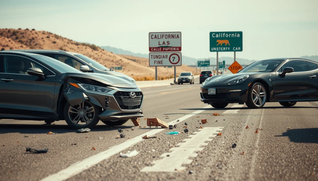
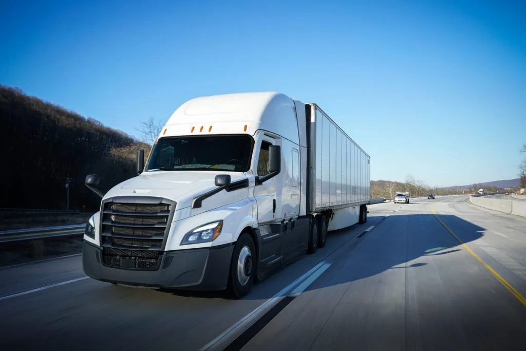
Motorcyclist Suffers Trauma in North Lakeport Major Injury Accident on S.R. 29 A North Lakeport major injury accident involving a truck and a motorcycle was…
Edward Smith
June 27, 2025
Posted In: Motorcycle Accident