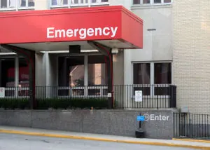
Prehospital and Hospital Evaluation of Chest Trauma
I’m Ed Smith, a Sacramento Trucking Accident Lawyer. While still in the field, the most that emergency medical technicians can do is to follow routine Advanced Trauma Life Support, which focuses on airway, breathing, and circulation. It is critical to take the patient to a high level trauma center as soon as possible, avoiding unnecessary interventions that may only delay definitive treatment.
Cervical spine immobilization should be done and high flow oxygen should be given by mask. The patient should be monitored via cardiac electrodes. There should be no delay in order to place IV lines or to intubate the patient unless the patient cannot be stabilized with a bag/mask device.
If there is no indication of respiratory difficulty or major injury, no intervention is likely to be necessary. The EMTs should, however, make note of the condition of the vehicle and steering wheel, whether or not the victim was ejected, significant intrusion into the passenger compartment, and whether or not there were fatalities among the other passengers. If the patient is hypotensive at the scene, this may indicate significant injury; this information should be passed on to the attending physician prior to arrival to the emergency department.
In the emergency department, airway, breathing, and circulation should be assessed via ATLS protocol. If, however, there is significant respiratory distress noted after chest trauma, breathing should be taken care of before the airway. If there is a tension pneumothorax inside the chest cavity, it should be relieved before doing the intubation. Any type of positive pressure ventilation will likely worsen a pneumothorax.
If the patient has unstable vital signs, low oxygen levels, or obvious major injury, the healthcare practitioner will perform a quick search and management of anything that is life threatening, including major injuries of the cervical spine, head, chest, pelvis, and abdomen.
Blunt chest trauma is associated with the following types of injurie besides cardiac contusion:
- Tension pneumothorax
- Aortic injuries
- Pericardial tamponade from a ruptured ventricular wall
- Blood in the chest cavity
- Disruption of the trachea or bronchi
Patients who are in significant respiratory distress, have a head injury, or have marked instability of vital signs should be intubated. Rapid sequence intubation is the preferable method, avoiding anything that causes further lowering of the blood pressure.
If there is a potential for a tension pneumothorax, a large needle should be used to decompress the pneumothorax. Needles may need to be as long as seven centimeters, depending on the size of the patient and should be placed in the second or third intercostal space or in the fifth intercostal space in the mid-axillary line. After that, a chest tube should be placed in order to keep the lungs expanded.
If the patient is stable in the emergency department and doesn’t need surgery, a CT scan of the chest with contrast should be done to identify which structures inside the chest are injured, particularly the aorta. If the patient cannot tolerate CT scanning because they need to go to surgery, a transesophageal echocardiogram can be done in the emergency department to assess the damage to the heart and aorta.
If the myocardium has ruptured from the injury to the heart, this can be seen as the presence of pericardial tamponade on ultrasound. If this is found, a pericardiocentesis can be done immediately to clear the blood from the pericardial sac. A catheter can be placed into this space to prevent re-accumulation of fluid, especially if transportation to another facility is indicated.
If there is still evidence of severe pericardial tamponade and percutaneous drainage does not work or the patient develops a cardiac arrest during resuscitation, a thoracotomy may have to be done in the emergency room to repair the cause of the bleeding.
If there is blood in the chest cavity, a chest tube should be placed in order to drain blood from this space. If there is immediate bloody drainage of at least 20 milliliters per kilogram, a thoracotomy must be done in the operating room.
In the event of blunt trauma to the chest, doing an emergency department thoracotomy rarely successfully resuscitates the patient. It works in only about five percent of patients who are already in shock and in about one percent of cases in which the patient has no vital signs. It never works in situations where there are no signs of life prior to transport to the hospital.
There are many risks to doing an emergency department thoracotomy and, for this reason, policies should be developed in order to determine when it should be done. Instead of a thoracotomy, the patient may need immediate surgery in order to live. Those who are likely to survive an emergency department thoracotomy include this subset of people:
- Patients who have suffered a cardiac tamponade that was quickly diagnosed and has no nonsurvivable injuries
- Patients who lose their vital signs in the emergency room and have no noted nonsurvivable injury
The procedure is likely to be futile if any one of these circumstances exist:
- The patient is not breathing, has no cardiac rhythm on EKG strip, and has no pulse
- The patient needed to have CPR for more than fifteen minutes prior to arrival to the emergency department
- The patient has any other nonsurvivable injury
There is some evidence to suggest that no patient with blunt trauma to the chest who needed more than five minutes of CPR at any time will survive with their brain function intact. A review of more than 25 studies involving more than 1300 patients who had sustained blunt trauma to the chest had a survival rate with an intact brain of only 1.5 percent. Only those who had measurable vital signs at the scene of the accident or in the emergency department and had a maximum duration of CPR of 11-15 minutes were able to survive.
Taking a history and monitoring the Patient
The first thing that must be done in any blunt thoracic injury is to quickly assess whether or not there has been injury to the heart, lung, or other mediastinal structures. A thorough history should be obtained if possible as well as a physical examination. If the patient is stable, x-rays, CT scans and echocardiography can be done to evaluate the possible damage to the heart.
The doctor should then be able to identify those patients who are at a high risk or low risk of major injury. The vital signs are the most important thing to assess as well as the patient’s clinical appearance. Remember that a young and healthy patient involved in a severe rollover accident may have no significant injuries, while at the same time, a frail, older patient who just tripped and fell may have multiple fractures of the rib and bruising of the lungs.
A general history and physical exam is generally not very sensitive for identifying injuries to the chest and hear. This is because these studies often do not pick up on the fact that the patient has sustained a cardiac injury and the follow up of these studies is poor.
Patients who are alert, in no discomfort, are not short of breath and have no tenderness to the chest wall are unlikely to have any serious blunt trauma injury. Those with low oxygen levels and abnormal chest sounds are usually the most specific for a hemothorax (blood in the chest cavity) or a pneumothorax. Chest tenderness and pain are sensitive indicators but re nonspecific.
A chest x-ray should be the first test done on all patients with blunt chest trauma. This is an inexpensive test that is both noninvasive and easy to get. In many cases, it can show useful information. For these reasons, a chest x-ray should be done on anyone who has sustained a blunt chest trauma, regardless of how minor the trauma should appear. A chest CT scan should follow as long as the patient doesn’t need to have surgery.
The chest x-ray can identify bruising of the lungs, a hemothorax and a pneumothorax. In some cases, it can show an injury to the aorta. The following findings on a chest x-ray should increase the chance that there is a blunt cardiac injury and should show which patients need further evaluation:
- Abnormal aortic shape
- Wide mediastinum
- Blood showing above the top of the left lung
- Large hemothorax on the left side
- A rightward deviation of the nasogastric tube
- Deviation of the trachea to the right side
A wide mediastinum is nonspecific but is otherwise considered sensitive for an injury to the aorta. These types of injuries account for about 20 percent of cases where there is a wide mediastinum after blunt injury to the chest. A CT scan of the chest may be done if the chest x-ray indicates the possibility of an aortic injury.
Those that deserve a portable chest x-ray include those who have a major mechanism of injury, those with low oxygen levels, those with abnormal vital signs, a positive seatbelt sight, evidence of multiple rib fractures, or significant tenderness of the chest. Those with an abnormal chest x-ray can go on to have a CT scan of the chest.
The patient who is stable with only minor abrasions, normal vitals, and mild tenderness can go to radiology for a regular PA and lateral chest x-ray. Those who are in pain and have tenderness of the ribs, chest pain on inspiration, or abdominal pain are at the highest risk of injuries to the chest and abdomen. Rib films are not usually necessary as they do not change the management of the patient.
The FAST examination (focused assessment with sonography in trauma) has become an important part of the evaluation of trauma patients, especially in patients with abdominal injuries and pericardial tamponade. Ultrasound of the chest is often done in the emergency department to check for hemothorax or pneumothorax.
The best test for blunt trauma of the chest is the CT scan of the chest with contrast. It shows the lung and mediastinal structures with a great deal of accuracy. It can identify a small pneumothorax, bruising of the lung, lacerations of the lung, and air in the mediastinum.
The use of CT of the chest in trauma patients has increased dramatically. In some places, CT is done even before a chest x-ray is done. Those with low risk mechanisms of injury, minor injury findings, and a normal chest x-ray usually don’t need to have a chest CT scan.
These people generally need a CT scan of the chest:
- Rapid deceleration injury
- Age older than 60 years
- Intoxication
- Pain in the chest
- Abnormal mental status
- Chest wall tenderness
- Another painful injury
If all of the above are absent, there is a low risk for chest injury and further imaging is not necessary. These guidelines can safely reduce the number of CT scans obtained, many of which are unnecessary. Even so, clinical judgement is necessary; if there have been any suspicious findings, a CT scan of the chest should be done.
I’m Ed Smith, a Sacramento Truck Accident Lawyer who has handled cardiac contusion injuries since 1982. Call me anytime for free, friendly advice at 916-921-6400 or outside Sacramento at 800-404-5400.
See our Reviews on Yelp, Avvo and Google.
Member of Million Dollar Advocates Forum.
See our past Verdicts and Settlements page.
Photo Attribution: Wikimedia Commons Public Domain Image Er.jpg

