

Traumatic Optic Neuropathy and Visual System Injury
Traumatic Optic Neuropathy and Visual System Injury
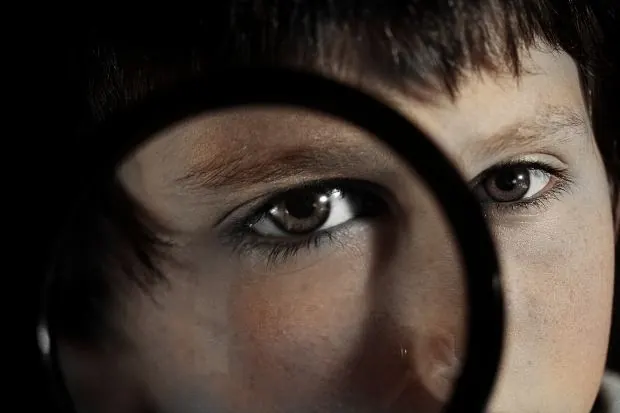
I’m Ed Smith, a Sacramento Eye Injury Lawyer. Eye lesions aren’t the only cause of impaired vision after trauma to the eye. The optic nerve, optic chiasm, and posterior visual pathways are all vulnerable in patients with open or closed head injuries. The ophthalmologist, focused on the care of the eye, must nevertheless be prepared to recognize and manage retrobulbar lesions.
One of the clinical advantages of ophthalmology is the ability to visualize most of the eye anatomy just by inspecting the globe. Unfortunately, with traumatic optic nerve and chiasmal lesions and abnormalities are apt to be situated beyond the reach of a flashlight, slit lamp, and ophthalmoscope. In cases of neural trauma, the ophthalmologist must be prepared to substitute inference for observation. Some neural lesions are missed because the ophthalmologist has simply failed to appreciate the possibility that the visual sensory pathway has been damaged behind the eye. This means that no assessment of the posttraumatic visual impairment can be considered complete unless the nerve and chiasm have been properly evaluated.
Ophthalmologists are usually forced to evaluate patients with visual loss immediately after head injuries and under difficult circumstances. The setting is often a hospital emergency department where special equipment is not available. It is also probable that the patient has other neurological or systemic injuries that make it complicated to evaluate and treat even the smallest eye injuries.
Since the care of the head injured patients with visual impairment is shared between ophthalmologists and others, it is essential that effective communication take place. This is accomplished usually through the medical record. Unfortunately, ophthalmologists often use arcane abbreviations and terms that other healthcare providers cannot interpret, even if the handwriting is legible. In the interest of good patient care, it makes sense to get rid of abbreviations and jargon so that the notes are legible and understandable to all.
Examination
Injuries to the retrobulbar visual pathways may have a lower priority when it comes to trauma but, when possible, alert and cooperative patients should be transported to a conventional eye examination facility if this doesn’t interfere with the care of their other traumatic injuries. If transport is not immediately available, the doctor should construct a preliminary examination at the bedside with plans for a more complete evaluation when the patient’s condition is better.
Acurate measurement of the patient’s visual acuity is the single most important aspect of the exam. In the setting of traumatic visual loss, with social and legal implications added to the medical complications, the visual acuity is one exam that the doctor should not delegate. The physician should do this evaluation him or herself.
If the patient is alert, ambulatory, and cooperative, the exam can be done easily using conventional measurements. Under the circumstances, however, improvisation may be required. Each eye must be tested individually, with one eye being patched at a time. Second, you should us a clean, well-illuminated chart and know the exact distance from the eye chart. This is sometimes a problem with the reduced Snellen charts (near charts) commonly used to test visual acuity in emergency departments and are designed to be used in patients in a stretcher. Some cards have a chain attached so that the exact distance to the chart can be determined. If not, you can easily attach a long string to the chart.
If the patient cannot see the largest Snellen chart better, it is better to move the chart closer to the patient and record the reading distance rather than use finger counting. Patient should wear their distance glasses if possible. If subnormal visual acuity cannot be obtained, a pinhole should be used to optimize the vision.
Color sense is particularly vulnerable to an injury to the optic nerve. This means that tests of color should be an essential part of the eye examination after traumatic visual loss. Ideally, one should use the pseudoisochromatic color plates designed for detecting congenital color blindness. Each eye should be tested individually and the test should be scored as the number of color plates detected correctly. When pseudoisochromatic plates are not available, a color comparison test can be used. The patient is asked to look at red bottle cap with each eye separately and is asked to compare the intensity of the color.
The pupillary evaluation is a key element in the recognition and identification of anterior visual pathway trauma. In patients unable to cooperate, such evaluation may be the only means of establishing the presence of an anterior visual pathway lesion. Pupillary size should be measured and not estimated; the direct and consensual reactions to light and near stimulation should be recorded.
The swinging flashlight test, in which a focused light is swung from one eye to the other, is especially helpful for identifying unilateral or asymmetric pupillary defects. A tendency for the pupils to dilate when the light is shone on one eye and to constrict when it is shown on the other eye indicates an afferent defect on the side that allows the pupil to dilate. If there is mydriasis from third nerve paralysis or other cause, one can still use the swinging flashlight test. One simply must observe the mobile pupil. A positive swinging flashlight sign in a patient with a normal fundus implies an injury to the retrobulbar area of the eye or a lesion of the anterior visual pathway.
The issue of instilling eye dilating drops for fundoscopy inevitably comes up in the course of evaluating head-injured patients with visual impairment. Certain simple rules should be scrupulously followed in this regard. Never dilate the pupils of an acutely injured patient with altered consciousness or one who is about to undergo a surgical procedure. Never dilate the pupils without the permission of the person’s primary physician. Make and observe your observations of the pupils before putting in the drops. Never use a long acting mydriatic. This means avoiding the use of scopolamine, atropine, and cyclopentolate. Instead use a drop such as tropicamide that rapidly produces adequate mydriasis of an acceptably short duration. Dilate both pupils at the same time. One dilated pupil is likely to be too confusing to other members of the team. Make a legible entry as to when you put the dilating drop into the eye.
Differential Diagnosis
There are many causes of visual loss after trauma and neural causes are much less common than eye causes. Therefore, it is important to look for signs of eye trauma as circumstances permit. Even if a retinal lesion is identified, one should always remain open to the possibility that a retrobulbar lesion also exists.
Missed intraocular lesions are by no means the only source of wrong diagnoses of traumatic neuropathies. Two other entities deserve consideration. The first is preexisting neuropathies and the other is factitious amblyopia. In such cases, optic atrophy will be evident at the time of injury. Since it takes weeks for optic atrophy to show up after retrobulbar trauma, the presence of optic atrophy immediately after the injury means that the optic neuropathy did not come from this trauma.
Factitious amblyopia must always be considered in the differential diagnosis of acute retrobulbar neuropathies since the combination of reduced visual function with a normal fundus is common to both. Some patients with factitious amblyopia ae malingering, while others have some psychiatric basis for their alleged visual deficits. It is not the ophthalmologist’s job to distinguish between the two.
Pupillary reactions may be helpful in differentiating factitious amblyopia from organic visual loss. Monocular or asymmetric visual impairment from a neural lesion should cause a positive swinging penlight sign. Factitious visual impairment does not alter the pupillary reactions. Visual fields may also be useful in diagnosing factitious amblyopia. Concentric field constriction is commonly encountered in these patients. A tubular configuration of the constricted field also strongly supports a diagnosis of factitious amblyopia.
The diagnosis of factitious amblyopia can be proven only by demonstrating that the patient can see better than they are alleging. This can be done using several techniques to fool the patient during the examination. The worse the alleged acuity, the easier it is to establish the diagnosis. Of course, patients may have both an organic visual defect and factitious amblyopia at the same time.
Direct optic nerve trauma refers to injuries resulting from objects penetrating the orbit and impinging upon the nerve. These injuries are encountered infrequently, and in virtually all that come to the attention of the ophthalmologist, the site of the injury is the intraorbital segment of the nerve. The intraorbital segment is lax and is therefore protected from all but high velocity impacts. Otherwise it would be hard to explain the few injuries seen among the thousands of patients who receive retrobulbar anesthetic blocks each year for ophthalmic surgery. One the other hand, high velocity impacts or any penetrating object that impinges on the nerve in the apex where it is tethered to the optic canal is likely to cause an optic neuropathy. Hemorrhages within and around the nerve are also common.
Indirect optic nerve trauma refers to the optic neuropathies that follow closed head trauma. Some experts divide these into an anterior and posterior variety. Anterior indirect injuries implicate the short intraocular segment of the optic nerve. The ophthalmological signs are invariably present in these types of injuries. Posterior indirect injuries refer to traumatic loss of vision that occurs without external or initial ophthalmological evidence of injury to the eye or to the nerve.
Anterior Indirect Neuropathies
Anterior indirect neuropathies are rare. Most cases occur when a blunt object intrudes between the globe and the orbital walls. Sometimes seemingly innocent injuries, such as getting poked while playing basketball can gouge the eye. At the other extreme are optic nerve avulsions self-inflicted by mentally ill patients. Anterior indirect optic nerve injuries happen when the globe is suddenly rotated or anteriorly displaced. This causes tears in the margins of the lamina cribrosa, which is the weakest portion of the ocular coat. The central retinal artery may be damaged in the process.
The management of these injuries is as follows: Patients with signs of central retinal artery occlusion deserve the same measures that are advisable for spontaneous central artery occlusions. Otherwise, provided there is no clinical or radiological evidence of another treatable lesion, special investigations or treatments are not warranted.
Posterior Indirect Optic Neuropathies
Posterior indirect neuropathies are seen rather more often than the direct or anterior indirect variety but they are by no means common. In contrast to indirect injuries that damage the intraorbital segment of the nerve and anterior indirect injuries that damage the intraocular segment, posterior indirect injuries typically damage the intracanalicular segment. This is the ten mm intracanalicular portion together with its complement of meninges, the ophthalmic artery, and sympathetic nerves crowded tightly within the bony canal behind the eye.
Many posterior indirect injuries appear to result from swelling and loss of blood supply to the affected area. Since swelling is treatable, this particular pathogenesis has important practical implications. Posterior indirect optic neuropathy usually involves young boys but can occur in both genders and at all ages. The injury involves blows to the face, forehead, or temple. The commonest mode of injury was a fall from a bicycle, closely followed by automobile accidents. The trauma may appear to be trivial. Nonvisual neurological deficits are seen among these patients with prolonged unconsciousness that is likely to be permanent. Some of these cases are bilateral.
Radiological testing is imperative whenever an optic nerve injury is considered in the pathogenesis of posttraumatic vision loss. CT scanning of the head and eyes without contrast injection is the initial study of choice. Bone window settings should be used to look for fractures. Neither the presence nor the location of the fracture correlated with the severity of the optic nerve damage.
The treatment is variable. Many patients resolve their visual acuity without intervention.
I’m Ed Smith, a Sacramento Eye Injury Lawyer. Call me anytime at 916-921-6400 or 800-404-5400 for free, friendly advice. Member of Million Dollar Advocates Forum.
Read our reviews on Yelp, Avvo and Google.