

Multiple Imaging Modalities: The Purpose of Each
I’m Ed Smith, a Sacramento Personal Injury Lawyer. Anyone who has been injured has likely been scanned with one or more types of imaging techniques. While these provide valuable pieces of information, each may play a significant role in the diagnosis of Reflex Sympathetic Dystrophy, also known as RSD.
Ultrasound for Fluid Pockets
First, if someone is involved in an auto accident and suffers traumatic injuries, they likely have received an ultrasound scan. Ultrasound is used to perform a FAST scan in the event of a traumatic accident. Meant to be performed rapidly, an ultrasound scan in this situation is used to look for fluid pockets in several different areas of the body that are commonly injured in traumatic accidents. If the patient’s extremities have been damaged, fluid can cause the hands and feet to swell. As a result, this swelling can stretch the nerves and produce the type of pain seen with RSD. Sometimes, this scan can reveal internal bleeding that requires emergent surgery.
X-Ray for Bone Fractures
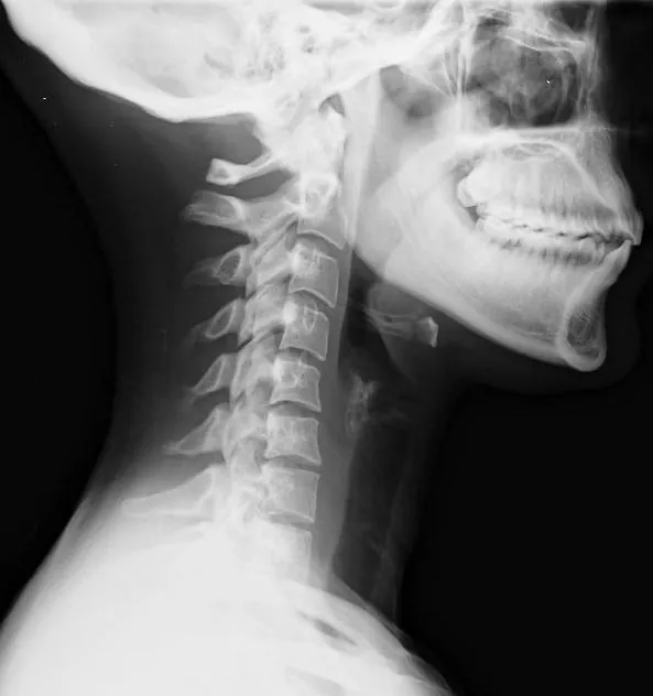
Bone fractures are a common occurrence in truck accidents and pedestrian accidents resulting in personal injury. An x-ray is used to detect fractures, such as femur fractures, because it is fast, safe, and cheap. Bone fractures in the patient’s extremities can lead to nerve damage that has been correlated with the development of RSD. If the patient has life-threatening injuries revealed on an x-ray, they will be taken for emergent surgery.
CT Scan for Bone Fractures and Bleeding
A CT scan is an imaging technique that uses a continuous x-ray scan that generates a 3D image. As a result, it provides more radiation but also more information. It can detect not only bone fractures but also bleeding. A CT scan used primarily to diagnose bone fractures and head injuries; however, the pain that patients complain about with RSD is transmitted to pain receptors in the patient’s brain. It’s not unusual for patients to receive brain scans if they are suffering from severe pain. Furthermore, if there is any concern for skull fractures or intracranial hemorrhaging, the patient will receive a CT scan.
MRI Scan is the Most Detailed
Next, an MRI is a magnetic scanner that provides a significant amount of detail and information. It uses magnets instead of radiation, making it a safe scan for anyone who doesn’t have any metal inside of their body; however, it also takes a long time. Because of this, an MRI is not commonly used in emergencies; however, it does provide important information regarding nerve function. It can be used in cases of RSD to examine the function of nerves in the extremities that may be responsible for producing severe pain.
Contact an Experienced Personal Injury Lawyer
I’m Ed Smith, a Sacramento Personal Injury Lawyer. Medical imaging is an essential part of proper diagnosis and treatment in Reflex Sympathetic Dystrophy. If you or a loved one has suffered an injury in an accident, please contact me at (916) 921-6400 for friendly, free advice. Anyone who is calling from outside of the local Sacramento area may want to use my toll-free number at (800) 404-5400.
I am a Million Dollar Advocate. This group is made up of trial lawyers who have obtained verdicts in excess $1 Million Dollars.
See the verdicts and settlements of our injury lawyers.
Comments from my past clients are available here: Avvo, Yelp, and Google.
Image Credit: Wikimedia Commons [public domain image]
:dr bw cv